
Medically reviewed by Dr. Ankur Gupta, BDS, MDS (Orthodontics) | Haryana Dental Council 1378A – Reg | 📅 Last medically reviewed:
Clinical Case Explainer · Zirconia Bridge & Emax Veneer · Loose, Gapped Front Teeth
A man in his mid-fifties had chipped, worn and widely spaced upper front teeth, and one root canal–treated tooth that had begun to loosen. This explainer walks through why the first plan of braces plus veneers was changed to a splinted zirconia bridge with an Emax veneer, and what decides between the two.
The Short Answer
When front teeth are chipped, worn and widely spaced, and one root canal–treated tooth has lost bone support, the choice is not made on appearance alone. It is made on how large the gap is and how stable the teeth are.
In this case, the first plan (braces to close the gap, then veneers) was changed to a 3-unit zirconia bridge on teeth 11, 21 and 22 with an Emax veneer on 12, because a loose tooth with bone loss may not suit being moved and then held on its own.
What the Patient Came In With
A man in his mid-fifties came in with one sentence: “Doc, I want to get something done about my front teeth.”
That sentence covered a lot. His upper front teeth were chipped, there were spaces between them, and some looked worn and stained. He also had a root canal-treated front tooth that no longer felt firm. None of this was an emergency, but each of these findings changed the plan.
What We Found on Examination
On clinical and radiographic examination:
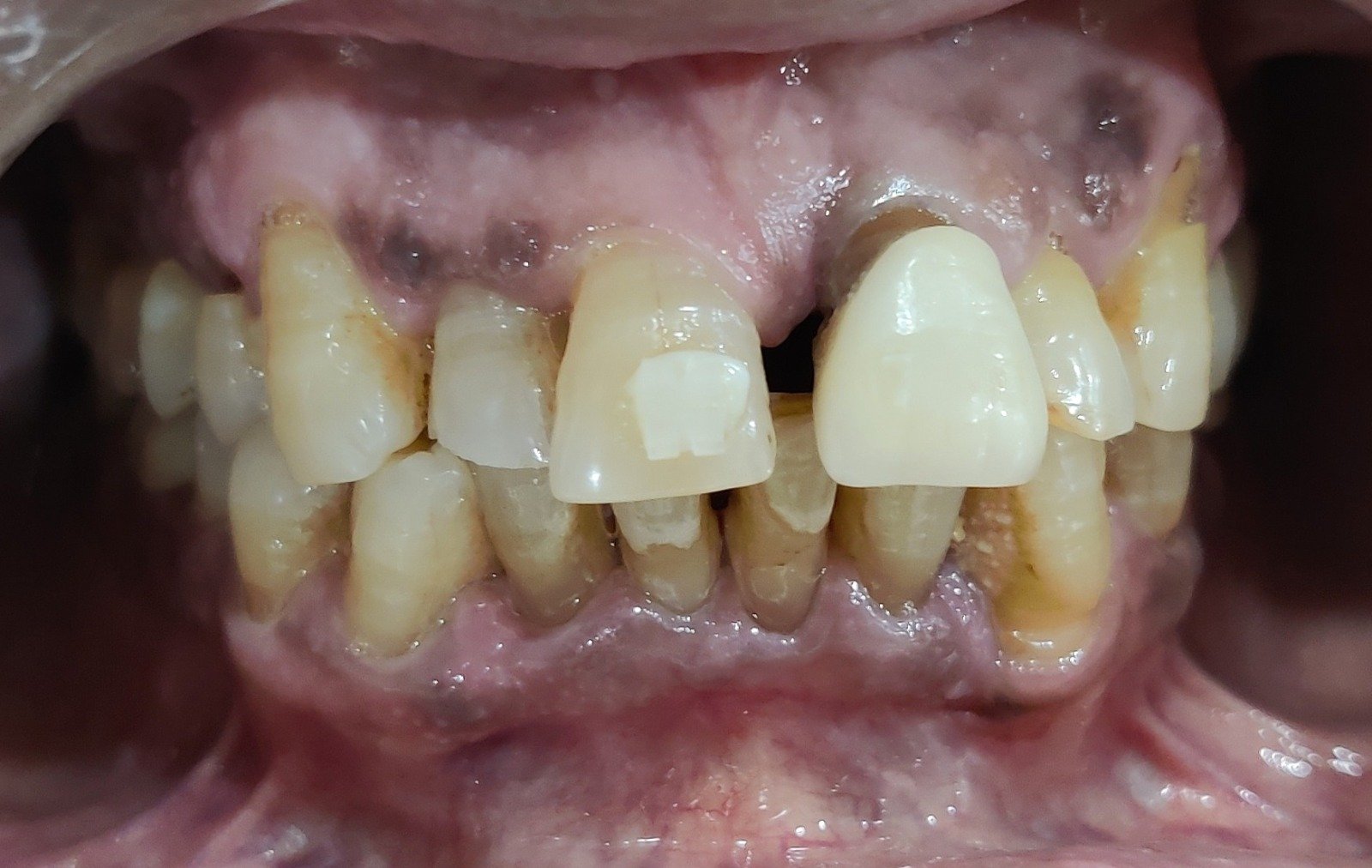
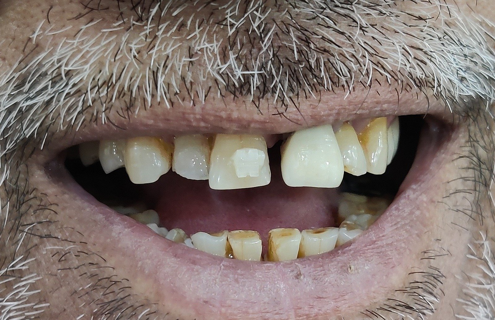
- Chipped upper front teeth with spacing between them, and a large gap between the two central incisors
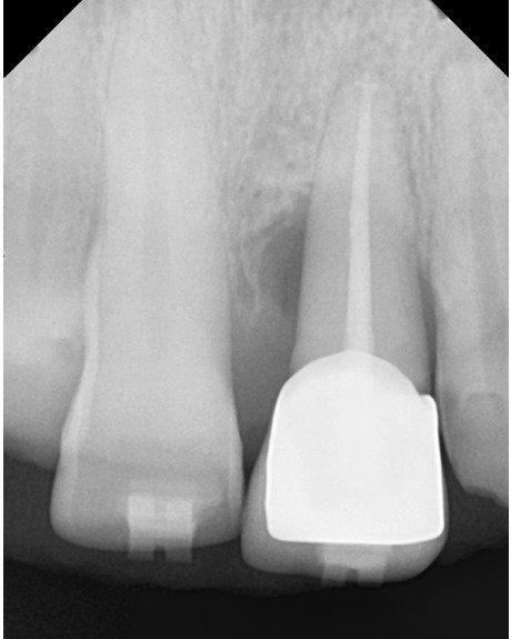
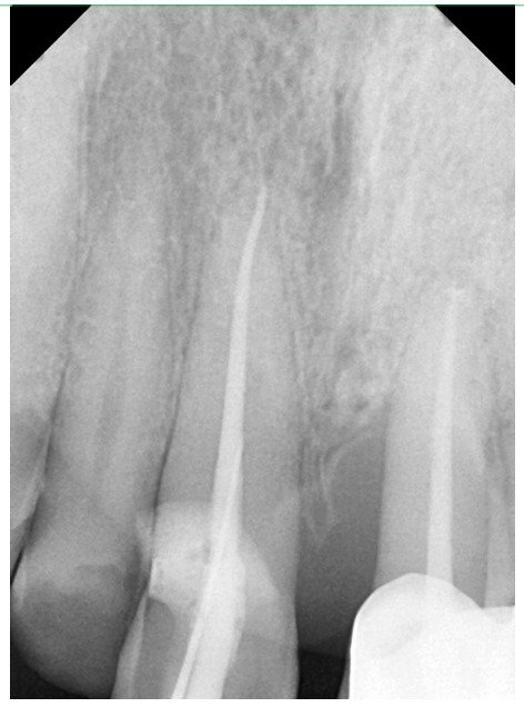
- Tooth 21: previously root canal–treated, with bone loss around it and Grade 1 mobility (a slight but measurable looseness)
- Tooth 12: worn down and discoloured, with similar wear on 11 and 22
- Teeth 11 and 22: sensitive to hot and cold
Every one of those findings affects which treatment can be offered. None of them can be judged from a photograph of a smile.
Why Not Simply Close the Gap and Add Veneers?
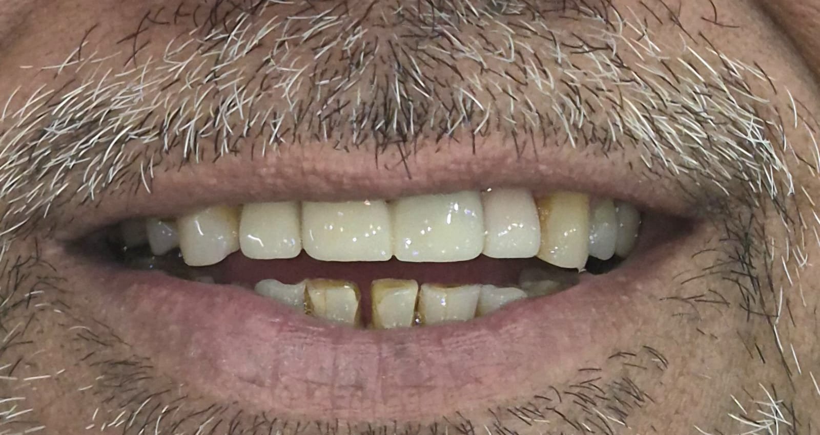
That was the first plan. Short-term braces on the front teeth would close the gap, and veneers on 12, 11 and 22 would improve shape and colour. A bonded attachment placed for this can be seen on one front tooth in the before-treatment photographs. Four things made us reconsider:
1. The size of the gap
The space between the central incisors was very large. Closing a large gap by tooth movement alone takes longer and asks more of the teeth, the gums and the patient than closing a small one.
2. The bone support around tooth 21
Teeth are moved through bone. Tooth 21 had bone loss and Grade 1 mobility, so it was not a tooth we could assume would tolerate being moved and then hold its new position by itself.
3. The condition of the neighbouring teeth
11 and 22 were sensitive to hot and cold, and 11, 22 and 12 were worn. Teeth in this state often need more than a thin veneer, and may need root canal treatment and full coverage.
4. Splinting
A fixed bridge joins neighbouring teeth into one unit. Joining a loose tooth to firmer neighbours lets them share the biting load. Whether this is suitable depends on the individual mouth.
The Two Options on the Table
| Option | Where it is strong | Where it is limited |
|---|---|---|
| Short-term braces on the front teeth, then veneers | Conserves tooth structure; moves teeth rather than covering the gap; suited to teeth with healthy bone support | Needs stable teeth and healthy bone; a large gap takes longer; a mobile tooth with bone loss may not tolerate movement; worn or sensitive teeth may still need coverage afterwards |
| 3-unit zirconia bridge on 11, 21, 22 with an Emax veneer on 12 | Rebuilds worn and chipped edges, closes the spacing and splints the loose tooth in one restoration; shorter active course | Requires reducing the teeth for crowns; needs root canal treatment where teeth are sensitive; the joined teeth must be cleaned carefully; restorations may need repair or replacement in time |
The plan was revised to the second option after the findings above were explained to the patient.
The Plan Agreed for This Patient
Phase 1 — Records and plan review
Examination, clinical photographs and X-rays, and a review of the first plan against the findings.
Phase 2 — Preparatory treatment
Root canal treatment of 11 and 22, which were sensitive to hot and cold. Deep gum cleaning (curettage) around 21, so that the gum around the loose tooth was as healthy as possible before the final restorations.
Phase 3 — Preparation and temporaries
11, 21 and 22 were prepared for the bridge and 12 for a veneer. Temporary restorations were placed in the same visit.
Phase 4 — Final fixing, 7 days later
The patient returned after 7 days. The 3-unit zirconia bridge over 11, 21 and 22 and the Emax veneer on 12 were fixed.
Phase 5 — Care and review
Crowns and bridges depend on how clean the gums stay. Regular cleaning, careful home care and routine check-ups are part of the plan, because no restoration lasts forever.
3-unit zirconia bridge (11 · 21 · 22)
Emax veneer
12
11
21
22
loose, bone loss
Patient’s right ← midline → Patient’s left (viewed from the front)
The sequencing principle, in one line: choose the treatment from what the examination shows (the size of the gap, the bone support and the condition of each tooth), not from the treatment the patient has heard of.
What Was Achieved
At the final visit the bridge on 11, 21 and 22 and the veneer on 12 were fixed. The chipped and worn edges were rebuilt, the spacing between the front teeth was closed, and tooth 21 was joined to its neighbours. At the final visit the patient reported that he was satisfied with how his front teeth now looked.
A note on this patient’s feedback: it is one person’s own account of how he felt about his appearance, shared without his name. It is not an endorsement and does not predict how anyone else will feel about their treatment. How any other mouth responds depends on its own bone support, gum health, bite and habits.
The lower front teeth were not part of this treatment.
What This Kind of Treatment Can and Cannot Do
| What it can do | What it does not do on its own |
|---|---|
| Rebuild chipped and worn front teeth and close spacing in one restoration | Treat active gum disease (gum health must be controlled first) |
| Join a loose tooth to its neighbours so they share the biting load | Rebuild lost bone or make a loose tooth firm again |
| Cover discoloured teeth with tooth-coloured ceramic | Remove the cause of the wear, such as grinding, if one is present |
| Be planned around teeth that have had root canal treatment | Last indefinitely (restorations can chip, loosen or need replacement) |
Signs It Is Worth Getting Your Front Teeth Assessed
- Front teeth that are chipping, or wearing flat or short
- A gap between the front teeth that has widened over time
- A front tooth that feels loose or moves when you bite
- A root canal–treated tooth that has darkened or feels different
- Teeth that are sensitive to hot or cold
- Bleeding or swollen gums around the front teeth
None of these is necessarily urgent, but each is easier to assess earlier than later.
Common Questions From Patients
What is a zirconia bridge?
A zirconia bridge is a tooth-coloured restoration made from a strong ceramic called zirconia. A 3-unit bridge joins three neighbouring teeth into one piece. In this case it covered 11, 21 and 22, so the middle tooth, which was slightly loose, was joined to its neighbours.
Why were three teeth joined together instead of treated one by one?
Tooth 21 had bone loss and Grade 1 mobility. Joining it to 11 and 22 lets the three teeth share the load of biting. Whether joining teeth is suitable depends on the bone support, the bite and the health of the gums, which is why it is decided only after examination.
Can braces close any gap between the front teeth?
Not always. Braces move teeth through bone, so the size of the gap, the health of the bone and gums, and the stability of each tooth all matter. A very large gap, or a loose tooth with bone loss, may be better managed another way, as it was here.
What is an Emax veneer?
An Emax veneer is a thin, tooth-coloured covering made from lithium disilicate ceramic, bonded to the front of a tooth to change its shape or colour. In this case one was used on 12, which was worn and discoloured.
Do I need a root canal before a crown?
Not always. Here, 11 and 22 were sensitive to hot and cold, so root canal treatment was done before the teeth were prepared. Whether a tooth needs it is decided after examining the tooth and taking an X-ray.
What are temporaries and why were they used?
Temporaries are short-term restorations that protect the prepared teeth while the final ones are made. In this case the patient wore them for 7 days before the final bridge and veneer were fixed.
How long will crowns and bridges last?
This varies from patient to patient and cannot be promised. It depends on gum health, cleaning, bite forces and habits such as grinding or clenching. Regular cleaning and check-ups are advised for any restoration.
How do I clean a bridge?
A bridge is cleaned like natural teeth, plus the area beneath and between the joined teeth, using floss threaders or interdental brushes. Your dentist or hygienist can show the method that suits your mouth.
What does it cost?
Fees depend on the number of teeth, the materials and any preparatory treatment such as root canals or gum treatment. A meaningful estimate is only possible after examination and X-rays, because a figure given before them is a guess.
What happens if I do nothing?
Chipped and worn teeth can continue to wear or break, and a loose tooth with bone loss may loosen further. Spacing can also widen over time. Not every case progresses, and a dental examination can tell you what is likely in yours.
Where We Are With This Case
The final bridge and veneer were fixed 7 days after tooth preparation. Review findings will be added to this page as they are recorded, so that the account reads as it happened rather than as a promise.
Patient details are anonymised. Clinical photographs and X-rays are published with the patient’s written consent, show teeth only, and are shared for education and awareness purposes only. Medically reviewed by Dr. Ankur Gupta, BDS, MDS (Orthodontics), Dr. Gupta’s Dental Implant & Orthodontic Centre. This article describes one patient’s presentation and treatment and is published for general education and awareness only. It is not a diagnosis and not a substitute for an in-person examination. Results vary from patient to patient and case to case, and nothing here is a guarantee or promise of any outcome. Only a dental consultation can decide what is the right treatment for you.